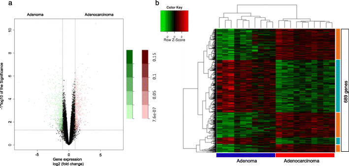
Fig. 1.
Microarray gene expression analysis in the 10 paired colorectal adenoma and adenocarcinoma samples of this study. A: Volcano Plot representing the set of genes analyzed by gene expression in the ten paired samples of colorectal adenoma and adenocarcinoma. Pink dots represent upregulated genes in adenocarcinoma when compared to adenoma; Green dots represent upregulated genes in adenoma when compared to adenocarcinoma. B: Unsupervised hierarchical clustering analysis in adenoma and adenocarcinoma samples based on the 689 differentially expressed genes (log 2 FC≥2 and FDR≤0,05)

