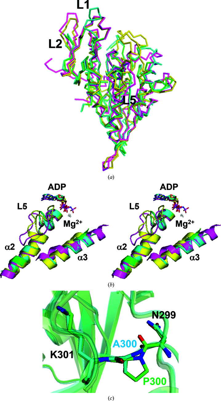
Figure 4.
Structural comparison with other structures. The structures of CENP-E–MgADP from this study (green), CENP-E–MgADP 1t5c (cyan), Eg5–MgADP (PDB entry 1ii6; magenta) and Eg5–MgAMPPNP (PDB entry 3hqd; yellow) and Mg2+ (white) are shown. (a) Ribbon representations of the structure of the CENP-E–MgADP from this study superposed with the previously reported structures of CENP-E–MgADP 1t5c, Eg5–MgADP (PDB entry 1ii6) and Eg5–MgAMPPNP (PDB entry 3hqd). The view is the same as in Fig. 1 ▸(a). (b) CENP-E has a unique structural orientation of L5 and helix α3 compared with ADP-bound and ATP-bound forms of Eg5 (stereoview). (c) Stick representations of superposed residues 299–301 of CENP-E–MgADP from this study and CENP-E–MgADP 1t5c.

