
Figure 2.
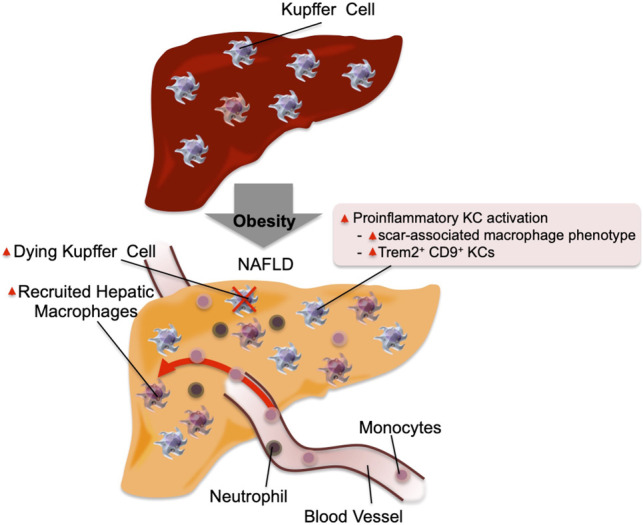
Inflammation in the liver. In the normal physiological state, KCs account for ∼10% of all liver cells. In addition, they scavenge pathogens and show M2-like polarized anti-inflammatory phenotype. In obese steatotic livers, KCs can show increased expression of genes associated with tissue repair and inflammation. These genes include Cd9 and Trem2. This is accompanied by increased KC apoptosis, and a component of KC death is compensated for by increased recruitment of blood monocytes, which differentiate into KC-like macrophages. Chemokines released by steatotic hepatocytes cause increased recruitment of blood monocytes into the liver, which differentiate into proinflammatory macrophages (RHMs) and produce factors that can cause insulin resistance. The recruitment of neutrophils also increases, and the molecules released from neutrophil granules such as neutrophil elastase and myeloperoxidase can induce insulin resistance in hepatocytes.