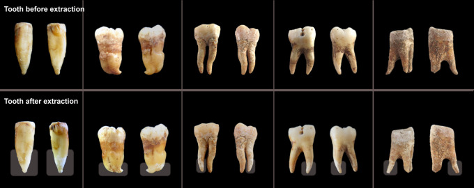
Figure 1.
Examples of teeth before and after minimally destructive extraction. Teeth which have been sampled using this minimally destructive extraction protocol were photographed prior to (top) and ∼24 h after (bottom) extraction. Through the use of parafilm to protect regions of the tooth that are not targeted during sampling, such as the crown, sample degradation is primarily restricted to the lower portion of the targeted tooth roots, and the overall morphology of the tooth remains intact. The region targeted for sampling (i.e., not covered by parafilm) is indicated by a transparent box in the after images. Note that these are representative examples of the typical impact of sampling using this method on ancient teeth of high quality (two left-most teeth) or moderate quality (three right-most teeth). Data from these teeth are not reported in this study. For before and after images of the tooth roots upon which sequencing was done during this study, see Supplemental Figure S1.

