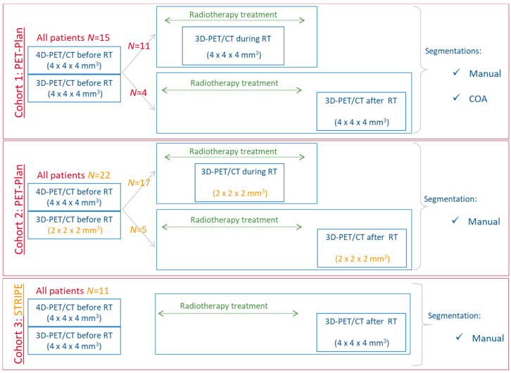
Figure 1.
Flowchart of the three cohorts: (i) cohort 1 and 3 were employed to identify the IF satisfying the criteria of being normally distributed across 4D PET and robust between 3D and 4D images; (ii) cohort 1 with manual segmentation of the primary tumor was the training cohort to develop the radiomics model for prediction of treatment response; and (iii) cohort 1 with COA (different segmentation) and cohort 2 (different voxel size for 3D image reconstruction and different patients) to validate the radiomics model.

