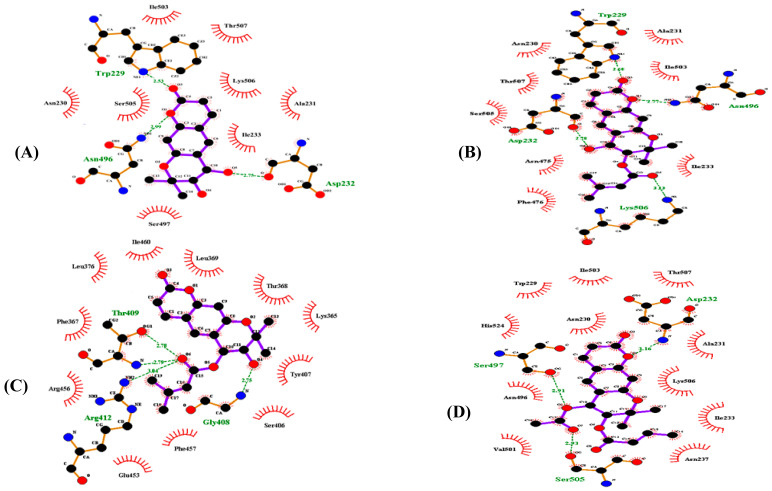
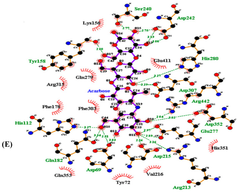
Figure 3.
Two-dimensional ligand interaction diagram of α-glucosidase inhibition by (+)-trans-decursidinol (A), Pd-C-I (B), Pd-C-II (C), Pd-C-III (D), and acarbose (E). Schematic display of the interaction of ligands (coumarins) and α-glucosidase, coumarins present thick purple stick models, hydrogen bonds are green dotted and hydrophobic interactions of dashed half-moons with the enzyme’s respective amino acid residue.


