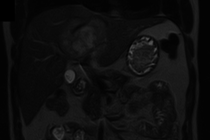
Figure 3.
An MRI (T2 weighted, T2W) abdomen and pelvis in the coronal plane, 17 days after presentation: there is a complex multiloculate lesion with restricted diffusion within segments IV and VIII, measuring 7.8x 5.9 cm in diameter. There are calculi within the gallbladder with no biliary dilatation.

