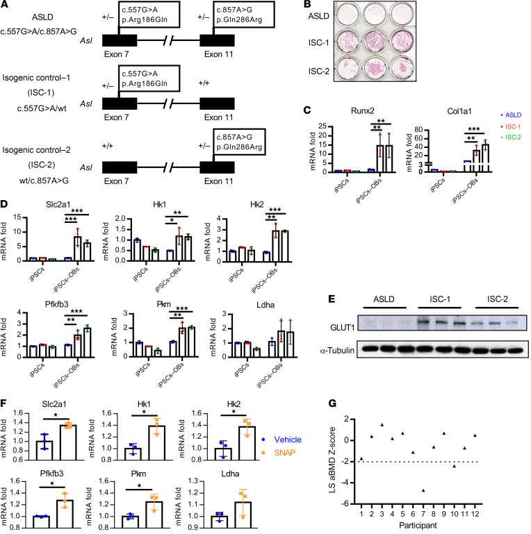
Figure 6. Effect of ASL deficiency on osteoblast function and bone mineral density in humans with ASLD.
(A) Schematic figure of correction of each of the variants of the compound heterozygous mutations of an individual with ASLD. (B) Alizarin red staining of ASLD and 2 isogenic control iPSC lines, differentiated in osteogenic medium for 21 days. n = 3. (C) mRNA levels of osteoblast marker genes Runx2, Col1a1, and (D) glycolytic genes Slc2a1, Hk1, Hk2, Pfkfb3, Pkm, and Ldha by qPCR, before (iPSCs) and after iPSCs were differentiated in osteogenic medium to osteoblasts (iPSCs→OBs) for 14 days. n = 3. (E) GLUT1 protein levels by Western blot. n = 3. iPSCs were differentiated in osteogenic medium for 14 days. n = 3. (F) mRNA levels of glycolytic genes Slc2a1, Hk1, Hk2, Pfkfb3, Pkm, and Ldha by qPCR. iPSCs from an individual with ASLD were differentiated in osteogenic medium and treated with 5 μM DMSO or 5 μM SNAP for 12 days. n = 3. Data are presented as mean ± SD. Student’s t test. *P < 0.05; **P < 0.01; ***P < 0.005. (G) Dot plot of aBMD z scores of lumbar spine (LS) regions L1–L4 in 12 individuals with ASLD. Two individuals had low bone density (L1–L4 z scores less than –2.0).

