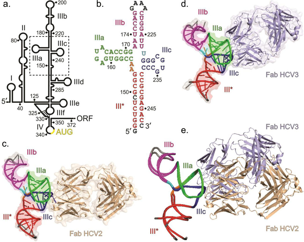
Fig. 1.
Overall structures of the HCV IRES JIIIabc in complex with Fabs HCV2 and HCV3. (a) Secondary structure of HCV IRES (genotype 1b) showing domains I – IV according to Brown et al.14 and Honda et al.16 Dotted box highlights the JIIIabc. Numbering depicts the approximate nucleotide position (b) The JIIIabc crystallization construct. Nucleotides in gray represent mutations or insertions compared to the wild-type (genotype 1b)13,15 sequence. (c) Crystal structure of the JIIIabc – HCV2 and (d) JIIIabc – HCV3 complexes solved at 1.81-Å and 2.75-Å resolution, respectively. (e) Superposition of the JIIIabc structures from JIIIabc – HCV2 and JIIIabc – HCV3 complexes. The JIIIabc structure is almost identical in both complexes; the HCV2 and HCV3 Fabs bind to the same region of the RNA with different orientations. Figures b-d and the corresponding labels are colored analogously for facile comparison.

