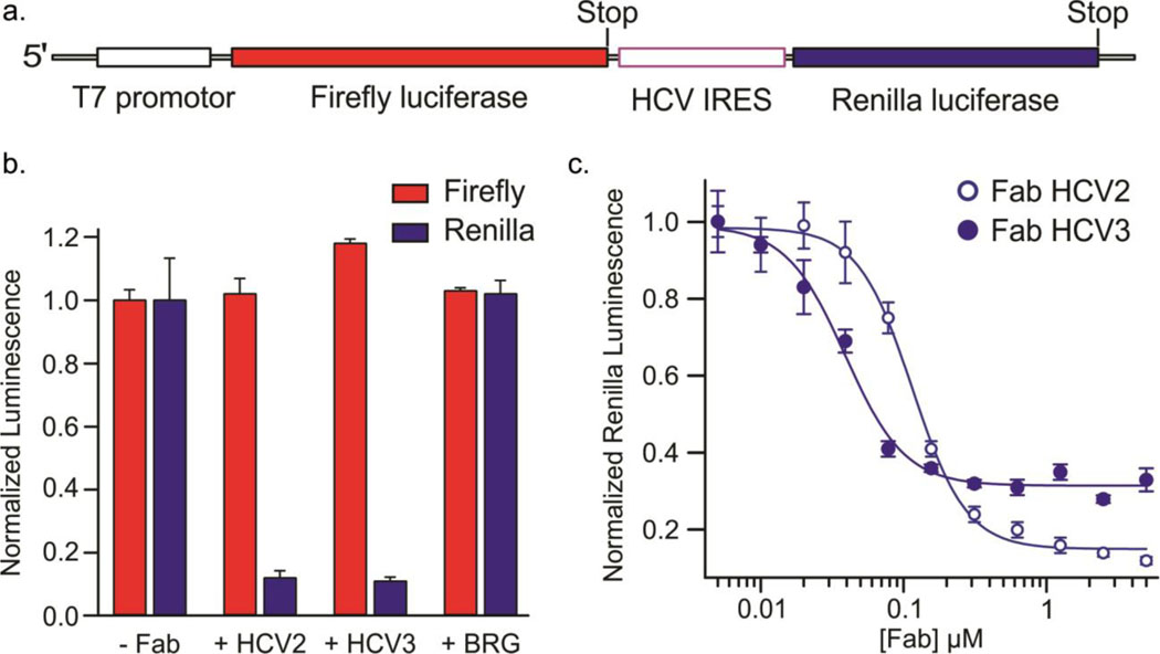
Fig. 5.
Inhibition of HCV IRES-mediated in vitro translation by Fabs HCV2 and HCV3 in a rabbit reticulocyte lysate-based assay. (a) Schematic of the dsDNA template used for generating the corresponding bicistronic mRNA for translation via a T7 promotor-controlled transcription. (b) Normalized luminescence corresponding to the expression of Firefly (red bars) and Renilla (blue bars) luciferases in the absence and presence of 5 μM of Fab HCV2, HCV3 or BRG. Fab BRG possesses the same scaffold domains as Fabs HCV2 and HCV3 but does not bind to the HCV IRES. (c) Normalized luminescence corresponding to the expression of Renilla luciferase with varying concentration of Fab HCV2 (solid circles) and HCV3 (hollow circles). Error bars in b and c represent the standard deviations from three independent experiments.

