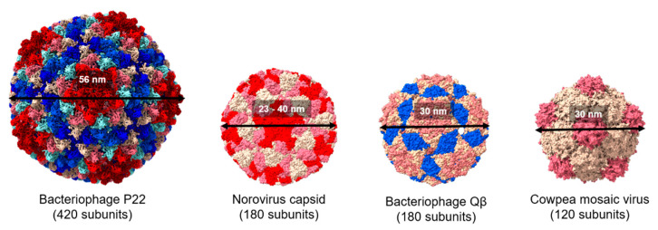
Figure 2.
Comparison of the structure of viral protein cages. Space filling models showing the structure of bacteriophage P22, norovirus capsid, cowpea mosaic virus, and bacteriophage Qβ (Protein Data Bank IDs: 2XYZ, 1IHM, 1NY7, and 1QBE, respectively). Subunit chains are distinguished by different colors. All the structures were generated using UCSF ChimeraX.

