Figure 11.
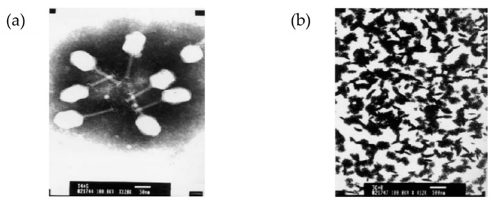
The head of bacteriophage T4 as a pathogenic display system. (a) Negatively stained image of the recombinant T4-3C phage particles (scale bar: 50 nm, magnification 12kx); (b) aggregation–precipitation of phages due to the addition of pig anti- foot-and-mouth disease (FMDV)-O antibody into the mixture of E. coli BL21 (DE3) infected phage T4-P1 and recombinant T4-3C phage particles (scale bar: 500 nm, magnification 12kx). Reprinted from [103], copyright (2008), with permission from Elsevier.

