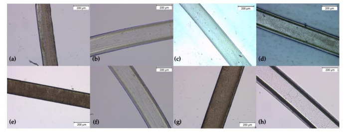
Figure 2.
Micrographs of the unloaded and loaded wet-spun fibers (a) SA, (b) SAGN, (c) SAGNCL and (d) GNCL, (e) SAz, (f) SAGNz, (g) SAGNCLz, (h) GNCLz, captured at 10× magnification using a brightfield microscope. The appearance of a coating-like layer surrounding the fibers is due to the microscope inability to perceive 3D structures, without losing focus.

