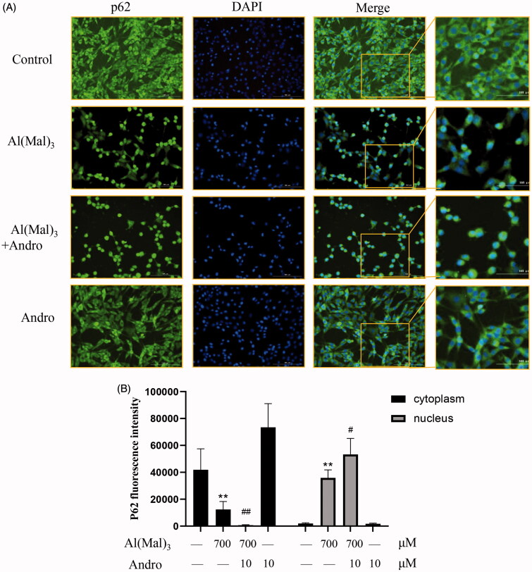
Figure 6.
Effect of Andro on expression and location of p62 protein in PC12 cells induced by Al(mal)3. Cells were incubated with 700 μM Al(mal)3 and 10 μM Andro for 24 h. The fluorescence localization of p62 was measured with immunofluorescence. An anti-p62 antibody was used to detect p62 localization using a fluorescence microscope. Green colour represented FITC-positive p62, Blue colour represented DAPI-positive nucleus. Bar = 100 μm (A); Quantitative immunofluorescence analysis of p62 in cytoplasm and the nucleus (B). *p < 0.05, **p < 0.01 versus the control, #p < 0.05, ##p < 0.01 versus Al(mal)3 group was considered statistically significant differences (n = 6).

