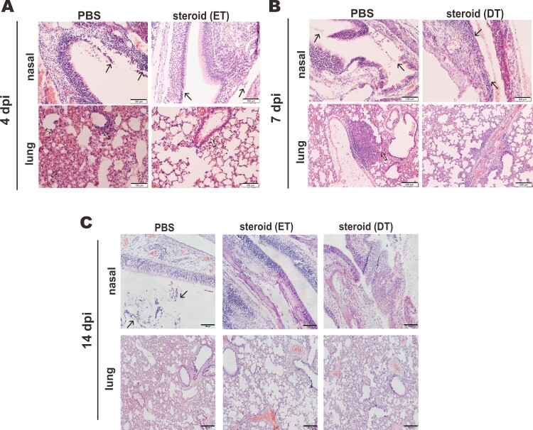
Figure 3.
Impact of steroid on the histopathological changes in nasal turbinates and lungs of SARS-CoV-2-infected hamsters. Representative images of nasal turbinate and lung sections of SARS-CoV-2-infected hamsters were shown. Hamsters were intranasally inoculated with 105 PFU of SARS-CoV-2 and then treated with methylprednisolone at 2 dpi (early steroid treatment, ET) or 5 dpi (delayed steroid treatment, DT). Histopathological changes in nasal turbinate and lung tissues were examined by hematoxylin and eosin staining. (A) Images at 4 dpi. Nasal turbinate tissue in PBS treatment control hamster showed severe epithelial desquamation and submucosal infiltration (arrows). The lung tissue showed diffuse alveolar wall thickening, blood vessel congestion, patchy area of alveolar space infiltration, exudation and patchy area of lung consolidation with two adjacent blood vessels showing vasculitis (open arrows). In ET group, respiratory and olfactory epithelium was intact, with mild submucosal infiltration observed in respiratory epithelium (arrows). In the lung tissue, only peribronchiolar infiltration and patchy area of alveolar wall thickening were observed (open arrows). Scale bar, 100 μm. (B) Images at 7 dpi showing histopathological changes in nasal turbinate and lung tissues from hamster receiving DT at 5 dpi. Nasal turbinate of the PBS control still showed some exudation mixed with cell debris in the nasal cavity (arrows), while the nasal epithelium showed prominent cell proliferation in steroid-treated hamster indicating tissue repairment (lower, arrows). Scale bar, 100 μm. The lung of PBS control hamster showed focal hemorrhage and patchy proliferative consolidation (open arrows); while in steroid-treated hamster, the lung still had mild perivascular infiltration but only very small foci of cell proliferation (open arrows). Scale bar, 200 μm. (C) Images at 14 dpi. Nasal turbinate tissue in PBS control hamster still showed mild degree intra-epithelium infiltration and submucosal blood vessel congestion. Luminal secretion with cell debris was occasionally observed (arrows). While nasal turbinate tissues showed intact epithelial layers, no apparent immune cell infiltration or luminal secretion was seen. Scale bar, 100 μm. The lung tissue in PBS control showed diffuse alveolar wall thickening and blood vessel congestion with no alveolar space infiltration or exudation. Upon early (ET) or delayed (DT) steroid treatment, the lung tissue showed only focal area of mild alveolar wall thickening and vessel congestion. Scale bar, 200 μm.

