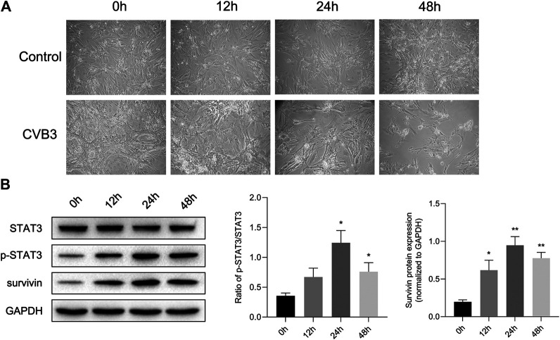
FIGURE 2.
STAT3 was activated in viral myocarditis cell model. NMCs were infected with CVB3 and the proteins were collected at different time point (A) The morphological changes of NMC at 0, 12, 24, and 48 h after CVB3 infection (200×) (B) The related proteins were measured by western blot (n = 4). *p < 0.05, **p < 0.01 vs control group (NC).

