Fig. 4.
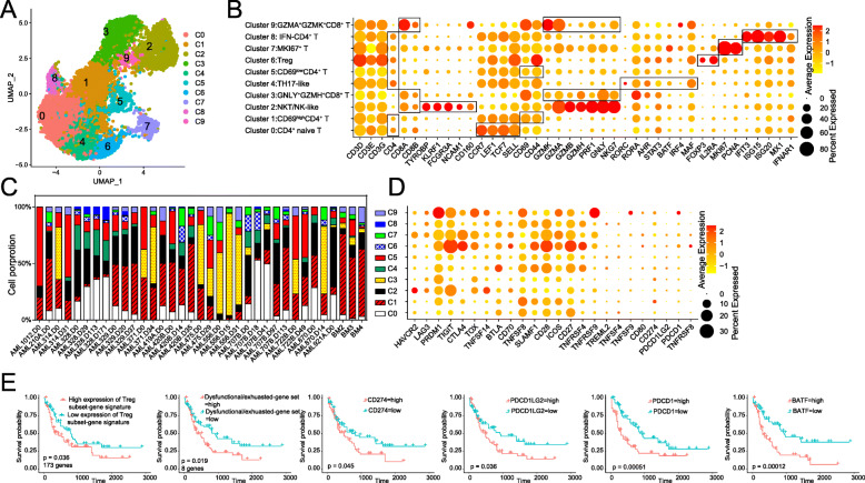
Diversity of T/NK subsets revealed by scRNA-seq analysis. a UMAP plot of sc-RNAseq data (n = 10,096 cells) showed 10 distinct clusters. b Dot plot of differently key cell-type marker genes. c Histogram showed the fractions of different cell-type in T/NK populations for each AML patient and healthy donors’ BM cells, colored based on cell type. d Dot plot showed the transcript expression pattern of stimulation molecules and their receptors. e The Kaplan-Meier overall survival curves of TCGA AML patients grouped by specific Treg gene sets, dysfunctional/exhausted-gene set (LAG3, TIGIT, CTLA4, HAVCR2, TOX, PDCD1, CD274, PDCD1LG2), and several genes (CD274, PDCD1LG2, and BATF). + represents censored observations, and P value was calculated by multivariate Cox regression

