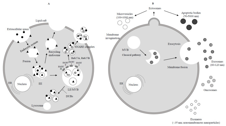
Figure 1.
Biogenesis of exosomes. (A) Endocytosis begins at the cytoplasmic membrane. Early endosomes (EEs) form by the fusion of uncoated endocytic vesicles. EEs either return to the plasma membrane (recycling endosome) or convert into late endosomes/multivesicular bodies (LEs/MVBs). Protein sorting of ILVs (intraluminal vesicles) occurs through endosomal sorting complexes required for transport (ESCRT) dependent or independent mechanism. ESCRT 0, ESCRT I, ESCRT II, ESCRT III are four components of ESCRT machinery that ubiquitinate the substrates on the part of the inward budding endosomal membrane. ILVs are ready to be degraded by lysosome or rescued by de-ubiquitinating enzymes (DUBs) through Rab GTPases (Rab27A and Rab27B) which allow the MVBs to move to the cell periphery. Finally MVBs through the SNARE complex fuse with the plasma membrane and lead to exocytosis (release of ILVs as exosomes to the extracellular space).  Extracellular protein;
  Receptor;
  Lipid;
  RNA. (B) Ectosomes derive from the plasma membrane by direct gemmation. Large oncosomes, as microvesicles originate from plasma membrane [8]. Exosomes are derived and secreted through a “classical” pathway. ER = endoplasmic reticulum.
  Microvesicle;
  Apoptotic bodies;
  Exosome;
  Exomere;
  Oncosome.

