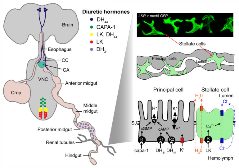
Figure 8.
Distribution and actions of LK and other diuretic hormones in adult Drosophila. A schematic depiction of the location of peptidergic neurons and gut endocrine cells that express diuretic hormones: capability-derived peptides (CAPA1-2), diuretic hormone 31 (DH31), diuretic hormone 44 (DH44) and leucokinin (LK). After release from these neurosecretory cells, the peptide hormones act on their receptors that are localized in either of two major cell types in the Malpighian (rental) tubules, the principal cells or stellate cells (visualized here using Lkr > mcd8GFP). The peptides act via different second messenger systems to alter the activity of ion pumps or channels. The orange rectangles represent aquaporin channels, the blue represent chloride channels and the red represents a Kir potassium channel. Abbreviations: CC, corpora cardiaca; CA, corpora allata; VNC, ventral nerve cord; SJ, septate junction; V, V-type ATPase. Figure 8 is based on a figure from [1]. The Malpighian tubule cell model is adapted and redrawn from O’Donnell et al. [131]. The image of localization of LKR in stellate cells is from Zandawala et al. [37] with permission from the publishers.

