

Abstract
Introduction
The posterior compartment of the thigh is composed of three major muscles collectively known as the hamstring muscles. These consist of the biceps femoris short and long head, semimembranosus and semitendinosus. Excluding the short head of biceps femoris, the hamstrings contribute to the movement of the hip and the knee joints as they span across both joints. Our hypothesis is that the nature of the conjoint tendon -ischial angle predisposes to an increased risk of tearing in this hamstring component. We therefore aim to look at the anatomy of the hamstring origin at the ischial tuberosity and spatial relationship between the long head of biceps, semitendinosus and semimembranosus in the form of vector angles.
Material and methods
100 consecutive pelvic MRIs in patients under the age of 40 years were reviewed by musculoskeletal radiology fellow and a consultant musculoskeletal radiologist with more than 10 years’ experience in musculoskeletal radiology and measured the angle of origin of conjoined tendon and semimembranosus at its ischial origin. P value using a paired t-test was less than 0.0001 confirming that the difference in the vector angle of the different hamstring components was statistically significant.
Results
The median angle of origin of conjoined tendon was 12° and for semimembranosus was 6°. Applying the concept of Newton’s second law to the angles calculated we demonstrated that an increase of 9% force applied to the conjoint tendon origin when compared to the semimembranosus tendon.
Conclusion
We hypothesis that the difference in the angle of origin of the components of hamstrings might be one of the reasons for the difference in the incidence and patterns of the injuries of the various muscles of the hamstrings.
Keywords: Hamstrings, Conjoint tendon, Semimembranosus, Vector angle
1. Introduction
The posterior compartment of the thigh is composed of three major muscles collectively referred to as the hamstring muscles. These consist of the biceps femoris short and long heads, semimembranosus and semitendinosus. Excluding the short head of biceps femoris, the hamstrings contribute to the movement of the hip and the knee joints as they span across both joints.1
This function renders this group of muscles essential to movement of standing, walking and running and therefore subsequently more vulnerable to injury. Our hypothesis is that the nature of the conjoint tendon -ischial angle predisposes to an increased risk of tearing in this hamstring component. We therefore aim to look at the anatomy of the hamstring origin at the ischial tuberosity and spatial relationship between the long head of biceps, semitendinosus and semimembranosus in the form of vector angles.
2. Background: anatomy of the proximal hamstrings
The biceps femoris muscle has two components, the long head arising from the medial facet of the ischial tuberosity and the short head arising from the lateral aspect of the linea aspera, lateral supracondylar line, and intermuscular septum.1
The semitendinosus and long head of biceps have a common origin from the ischial tuberosity, whereas the semimembranosus has a separate origin. The semimembranosus footprint on the ischial tuberosity lies anterior to that of the conjoint tendon. The semimembranosus also has a longer footprint than that of the conjoint tendon.2 Different authors have described the ischial origin differently. Battermann et al. for instance describes three different facets of the ischial tuberosity. The semitendinosus arising inferiorly with the main portion of the tendon arising from a medial facet together with the biceps femoris. Semimembranosus however arises from the lateral facet.3
Hamstring muscle injury commonly occurs at the musculotendinous junction. A study by de Smet et al. states that biceps femoris is the most commonly injured hamstring muscle. Semitendinosus is the second most commonly injured muscle followed by semimembranosus.4
Made et al. suggest that the architectural characteristics of the hamstring muscle complex may play a role in hamstring injury.5
While the authors look at the length of the different tendons and the musculotendinous junction, the angle of origin of the hamstrings has not been previously examined.5 We have studied the angle of origin of the different hamstrings and have extrapolated the force across each tendon. This is not a definitive study rather a suggestion that should lead to further research on the biomechanics of the hamstrings.
3. Methods
This was a retrospective study in which 100 consecutive pelvic MRI’s in patients under the age of 40 years (age range 18–40) with a male predominance (M:F, 72:28) were reviewed. The clinical indication for the MRI was either groin or hip pain. Images were reviewed by a musculoskeletal radiology fellow and a consultant musculoskeletal radiologist with more than 10 years’ experience in musculoskeletal radiology. Patients with a history of previous surgery in the pelvis or metalwork in the proximal femur and pelvis were excluded from the study.
The angle of origin of the hamstring components was then measured. Using a coronal non-fat saturated T1 sequence mid-hamstring image a line was drawn from the ischial hamstring origins (line x). A line (line y) was then drawn perpendicular to the first line (line x), (Fig. 1). The perpendicular line (line y) was then used as a reference for the best-fit angle for the conjoint tendon origin and separately the semimembranosus. (Fig. 2, Fig. 3, Fig. 4).
Fig. 1.
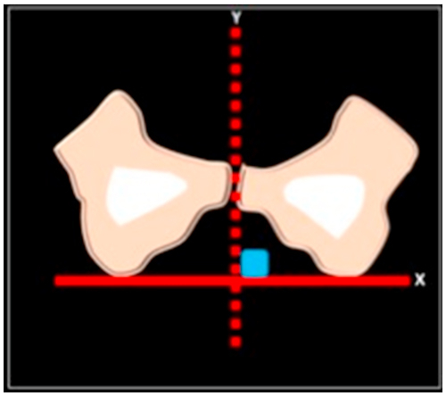
Line (y) drawn perpendicular to the inter-ischial line (x).
Fig. 2.
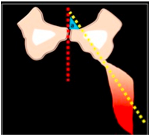
Angle of origin for conjoint tendon (angle a).
Fig. 3.
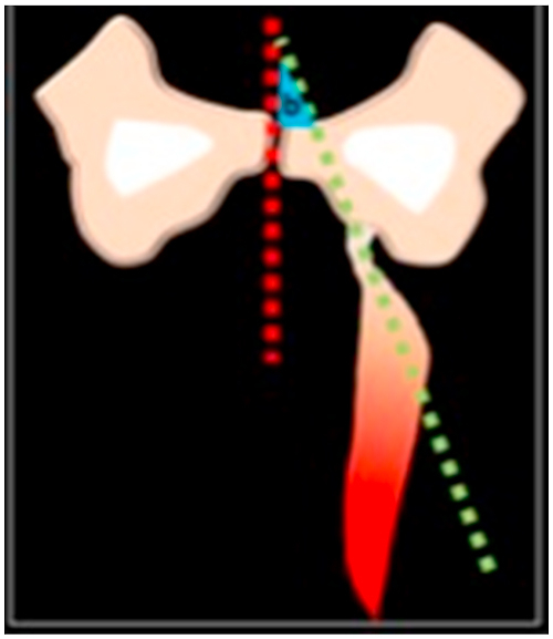
Angle of origin of the semimembranosus tendon (angle b).
Fig. 4.

Coronal T1 non-fat sat image at the mid hamstring plane with a trans-ischial line (x) and a perpendicular line (y) drawn. The angle of origin for the conjoint tendon (a) and semimembranosus (b) were subsequently extrapolated.
The mean, median, range and p values (using 202 measurements for 100 patients, with no exclusions) were calculated using a paired t-test.
The force value of each of the individual hamstrings was extrapolated from the value of the different vector angles of the individual hamstring components similar to descried by Guex and colleagues. This was generated using the concept of Newton’s second law. This states that the acceleration of an object as produced by a net force is directly proportional to the magnitude of the net force, in the same direction as the net force, and inversely proportional to the mass of the object.7 Using this concept we suggest this equation:
where F∗ is the force required if the tendon were vertical (theta = 0°).
4. Results
The median value of the vector angles for the conjoined tendon (long head of biceps femoris and semitendinosus) is 12° (range is between 6 and 21) and that of the semimembranosus is 6° (range is between 1 and 13). There was a good interobserver reliability. P value using a paired t-test was less than 0.0001 confirming that the difference in the vector angle of the different hamstring components was statistically significant.
Applying the concept of Newton’s second law as described above, we observed that a force of 1.10F (10% excess force) was needed for an angle of 6° (semimembranosus tendon origin) and 1.19F (19% excess force) for 12° angle (conjoint tendon origin) demonstrating an increase of 9% force applied to the conjoint tendon origin when compared to the semimembranosus tendon (Table 1).
Table 1.
Angle in degrees of origin of components of hamstrings.
| Conjoined tendon | Semimembranosus | |
|---|---|---|
| Median | 12 | 6 |
| Range | 6 to 21 | 1 to 13 |
| Mean | 12.33 | 5.07 |
The forces of the semimembranosus and the conjoint tendon are derived from the angles we calculated using equation F = F∗/cos (theta).
5. Discussion
Hamstring injury is often caused by sprinting or jumping. This is frequently seen in athletes playing soccer, rugby and basketball.8 Hamstring injuries are most common sports injury and can be associated with long rehabilitation period and recurrence. MRI has been used to diagnose and grade hamstring injuries and also to evaluate the prognosis of muscle tears and its impact on the time to return to play.
Few studies have used routine MRI images to calculate the implication of hamstring anatomy on the inherent risk of tears.
In our study we postulate that as the angle of origin of the conjoint tendon and the semimembranosus tendon is different this may subsequently affect the force exerted on the tendon during muscle contraction. The difference in the angles of origin of the different hamstrings was statistically significant (p < 0.05). There are however other confounding factors, which may influence the force, exerted by the hamstring rather than the angle of origin alone. This would affect the force values resulting from the angles calculated in this study. Further biomechanical studies may be needed which would take into consideration other factors such as muscle cross-sectional area and muscle fibre type.9
A force acting at an angle can be split into perpendicular components or angles, applying Newton’s second law to each of the perpendicular components then translates into the force applied at the hamstring origin. We used this concept to determine the effect of the angle of origin of the hamstrings on the resulting force applied to the individual components of the hamstring compartment. The forces applied are higher in the conjoint tendon and we propose that this may be one of the underlying factors related to the propensity of this hamstring component to tear.
A large retrospective observational study by Crema et al. has shown that the more commonly injured hamstring muscle is the long head of biceps and this tends to occur in a more proximal location.10
Myotendinous hamstring injuries are often caused by indirect trauma usually resulting from excessive stretching or tension and eccentric exercise result in lengthening of the contracting muscle. This may then result in microscopic damage in the muscle fibres or strain-type injury9. There are multiple hypotheses suggested as the underlying cause for biceps femoris being the more commonly injured hamstring. Bencardino et al. attribute the limited extensibility of the muscle, which originates from the femur as a contributing factor whilst Smet and Best also suggest that the type of sport played plays an important contributing factor as demonstrated in their series as all athletes with isolated semitendinosus injury were track and field jumpers.11,12
Further studies of the biomechanics and isokinetics of the hamstring muscle complex would be able to delineate the other confounding factors, which make the biceps femoris and conjoint tendon most likely to be injured. Our theory may be confirmed by being applied to biomechanical models.
References
- 1.Vaughn J.E., Cohen-Levy W.B. Anatomy, bony pelvis and lower limb, posterior thigh muscles. StatPearls. 2019 Treasure Island (FL): StatPearls Publishing; May 20. [PubMed] [Google Scholar]
- 2.Feucht M.J., Plath J.E., Seppel G., Hinterwimmer S., Imhoff A.B., Brucker P.U. Gross anatomical and dimensional characteristics of the proximal hamstring origin. Knee Surg Sports Traumatol Arthrosc. 2015;23(9):2576–2582. doi: 10.1007/s00167-014-3124-0. [DOI] [PubMed] [Google Scholar]
- 3.Battermann N., Appell H.J., Dargel J., Koebke J. An anatomical study of the proximal hamstring muscle complex to elucidate muscle strains in this region. Int J Sports Med. 2011;32(3):211–215. doi: 10.1055/s-0030-1268011. [DOI] [PubMed] [Google Scholar]
- 4.De Smet A.A., Best T.M. MR imaging of the distribution and location of acute hamstring injuries in athletes. Am J Roentgenol. 2000;174(2):393–399. doi: 10.2214/ajr.174.2.1740393. [DOI] [PubMed] [Google Scholar]
- 5.van der Made A.D., Wieldraaijer T., Kerkhoffs G.M. The hamstring muscle complex. Knee Surg Sports Traumatol Arthrosc. 2015;23(7):2115–2122. doi: 10.1007/s00167-013-2744-0. [DOI] [PubMed] [Google Scholar]
- 7.Guex K., Gojanovic B., Millet G.P. Influence of hip-flexion angle on hamstrings isokinetic activity in sprinters. J Athl Train. 2012;47(4):390-395. doi: 10.4085/1062-6050-47.4.04. [DOI] [PMC free article] [PubMed] [Google Scholar]
- 8.https://www.physicsclassroom.com/class/newtlaws/Lesson-3/Newton-s-Second-Law
- 9.Slavotinek J.P., Verrall G.M., Fon G.T. Hamstring injury in athletes: using MR imaging measurements to compare extent of muscle injury with amount of time lost from competition. Am J Roentgenol. 2002;179(6):1621–1628. doi: 10.2214/ajr.179.6.1791621. [DOI] [PubMed] [Google Scholar]
- 10.Fitts R.H., McDonald K.S., Schluter J.M. The determinants of skeletal muscle force and power: their adaptability with changes in activity pattern. J Biomech. 1991;24(SUPPL. 1):111–122. doi: 10.1016/0021-9290(91)90382-w. [DOI] [PubMed] [Google Scholar]
- 11.Crema M.D., Guermazi A., Tol J.L., Niu J., Hamilton B., Roemer F.W. Acute hamstring injury in football players: association between anatomical location and extent of injury-A large single-center MRI report. J Sci Med Sport. 2016;19(4):317–322. doi: 10.1016/j.jsams.2015.04.005. [DOI] [PubMed] [Google Scholar]
- 12.Bencardino J.T., Mellado J.M. Hamstring injuries of the hip. Magn Reson Imag Clin N Am. 2005;13(4):677–690. doi: 10.1016/j.mric.2005.08.002. [DOI] [PubMed] [Google Scholar]