
Figure 2.
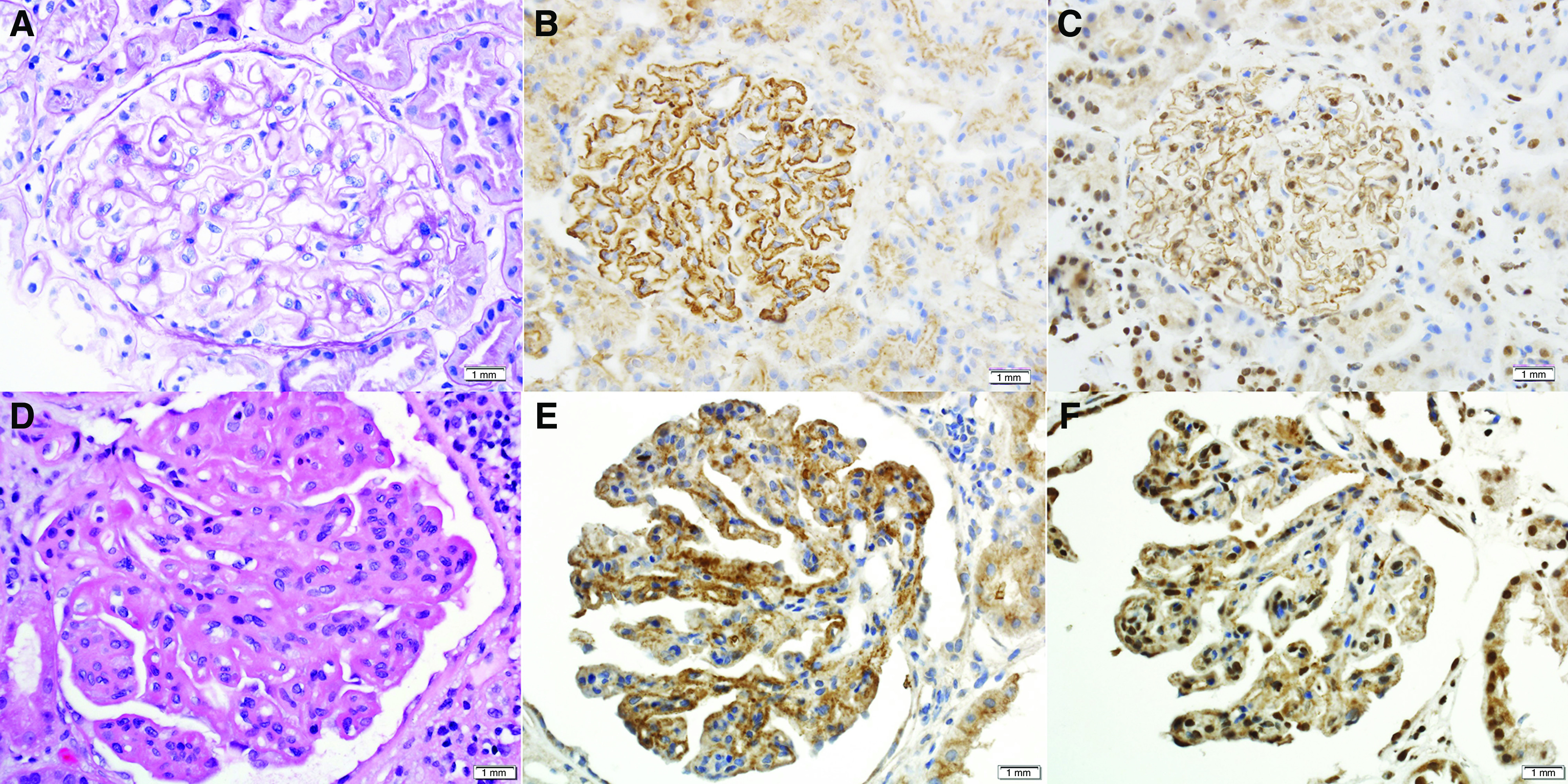
Light microscopy and immunohistochemistry (IHC) of EXT1/EXT2-positive LMN.(A–C) Top panel showing pure class 5 LMN, and (D–F) bottom panel showing class 5 LMN with class 3 proliferative LN. (B and E) IHC for EXT1 (×40 magnification). (C and F) IHC for EXT2 (×40 magnification).