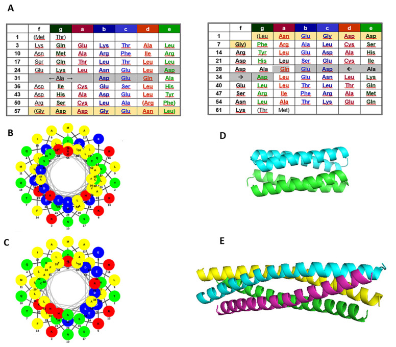
Figure 1.
Sequences and available structural data of the proteins examined. (A) Rop and RM6 sequences with the assignment of heptad positions (left), rRop and rRM6 sequences with heptad positions assigned in analogy to their parent proteins (right). RM6 and rRM6 lack the residues with grey shading. Light yellow shading indicates disordered residues in Rop and RM6, which do not participate in the coiled-coil structure. Similar shading has been used for their counterparts rRop and rRM6. (B,C) Helical wheels representation of Rop (B) and RM6 (C), with polar residues displayed with red (basic), blue (acidic), and green (uncharged) circles, and non-polar residues with yellow. (D) Structure of the Rop dimer (PDB id 1ROP). (E) Structure of the RM6 tetramer (PDB id 1QX8).

