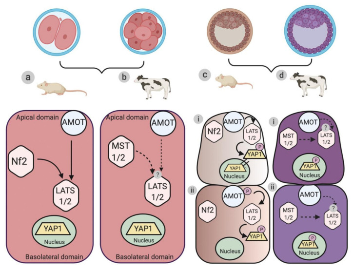
Figure 1.

Schematic illustration of the differences in the localization of the upstream regulators and the core cascade components of the Hippo signaling pathway during murine and bovine embryogenesis. (a) Organization hierarchy of the upstream regulators (AMOT and Nf2) and core cascade components (large tumor suppressor 1 and 2 (LATS1/2) protein kinase) of the Hippo signaling pathway during pre-compaction stages (two-cell to eight-cell stages) of mouse embryogenesis. Arrows represent the direction of activation of the protein kinase components. (b) Protein localization of the upstream regulators (AMOT and Nf2) and the core cascade components (mammalian sterile twenty like 1 and 2 (MST1/2) and large tumor suppressor 1 and 2 (LATS1/2)) of the Hippo signaling pathway during the pre-compaction stages of bovine embryogenesis. Dotted arrows represent a potential link in the activation of the protein kinase components. (c,d) Localization of AMOT, Nf2, MST1/2, and LATS1/2 in TE (trophectoderm) and ICM (inner cell mass) during blastocyst formation in murine and bovine models, respectively. (c-i) The Hippo signaling pathway is inactive (Hippo “Off”) in the outer polar TE cells, where AMOT and Nf2 cause the nuclear retention of YAP1. (c-ii) The Hippo signaling pathway is active (Hippo “On”) in the apolar ICM cells, where AMOT and Nf2 cause the phosphorylation of LATS1/2 and the subsequent cytoplasmic retention of p-YAP1. (d-i and ii) Protein localization of upstream regulators (AMOT) and core cascade components (MST1/2 and LATS1/2) of Hippo signaling pathway components in TE and ICM during bovine blastocyst formation. Dotted lines represent the proposed mechanism of functioning of the Hippo signaling pathway during (i) TE and (ii) ICM formation.
