Figure 1.
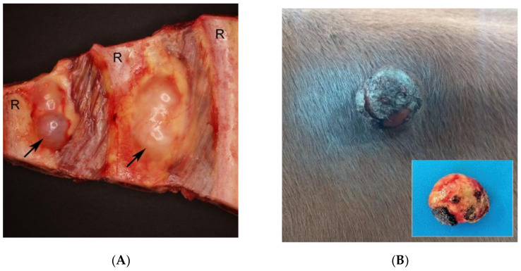
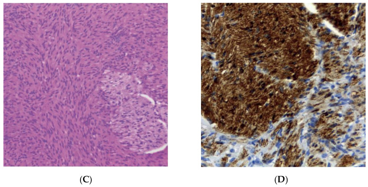
Spontaneous features of neurofibromatosis (NF) in cows. Bovine peripheral nerve sheath tumor (PNST) (A) and cutaneous neurofibroma (B–D). (A) PNST. Two tumors (arrows) located in relation to intercostal nerves (not visible). Specimen displaying the internal surface of the thorax. R: rib [42]. (B) Cutaneous neurofibroma. Note the firm exophytic nodule with marked ulceration in the paralumbar fossa. Inset: Nodule after complete surgical excision [54]. (C) Cutaneous neurofibroma. Note the mesenchymal cell proliferation forming plexiform beams. Hematoxylin-Eosin. ×200 [54]. (D) Cutaneous neurofibroma. Positive immunoblotting for S-100, counterstained with Harris Hematoxylin. ×400 [54].


