Figure 2.
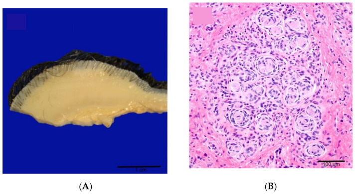
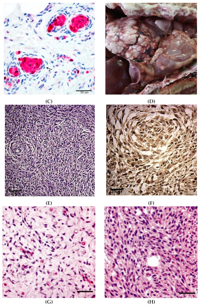
Spontaneous features of NF in dogs. Cutaneous neurofibroma (A–C), malignant peripheral nerve sheath tumors (MPNST) (D–F), and malignant transformation to MPNST (G-H). (A) Gross appearance of a neurofibroma effacing the dermis and subcutis of haired skin [20]. (B). Tactile-like structures characterized by parallel stacks or whorls 3–7 cells thick surrounded by a perineurial cell capsule (pseudomeissnerian corpuscles) in cutaneous neurofibroma. H&E [20]. (C) S100 immunohistochemistry (IHC) of cutaneous neurofibroma reveals positive staining of the pseudomeissnerian corpuscles (star) but lack of stain uptake in the perineurial cells (arrow) [20]. (D) Pulmonary MPNST in Cocker Spaniel dog with concomitant cNF. White multilobular mass in the caudal right lung [61]. (E). Section from the pulmonary mass showing dense proliferation of neoplastic cells arranged in interwoven bundles and concentric whorls (Antoni A pattern). H&E [61]. (F). Section from the pulmonary mass showing strong expression of S100 IHC [61]. (G) Golden Retriever dog with right hind limb enlargement. The sciatic nerve is infiltrated by a hypocellular well-differentiated spindle cell neoplasm diagnosed as benign PNST. Bar = 100μm [64]. (H) The large soft tissue mass surrounding the right ischium is composed of sheets of malignant spindle cells. Bar = 100 μm [64].


