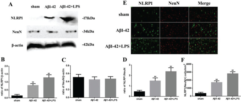
Figure 1. (A) Western blotting assay for detecting the expression of NLRP1 and NeuN. (B, C) Quantification of the expression of NLRP1 and NeuN. (D) NLRP1/NeuN ratio. (E) Immunofluorescence assay for detecting the expression of NLRP1 and NeuN (×400) and (F) NLRP1/NeuN (+) cells in samples from the sham, Aβ1-42, and Aβ1-42+LPS groups. Protein levels were normalized to those of β-actin (Aβ1-42 vs. sham group, **p<0.05; Aβ1-42+LPS vs. Aβ1-4 group, ## p<0.05, n=6 per group. All data were represented as the mean±standard error).

