Figure 6.
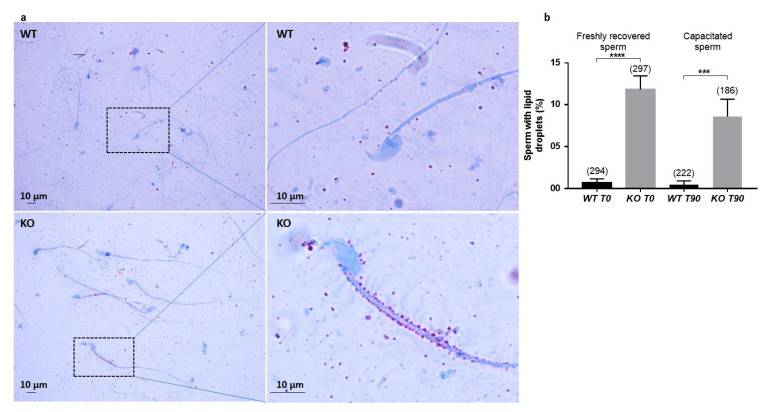
Lipid detection by Oil Red O staining. (a). Light microscopy detection of lipid droplets after Oil Red O staining. Right panels show higher magnification of regions framed in left panels. (b). Counting of sperm positive for lipid droplets after staining, before (T0) or after (T90) capacitation. Numbers between brackets indicate the counted sperm; *** p < 0.001 **** p < 0.0001.

