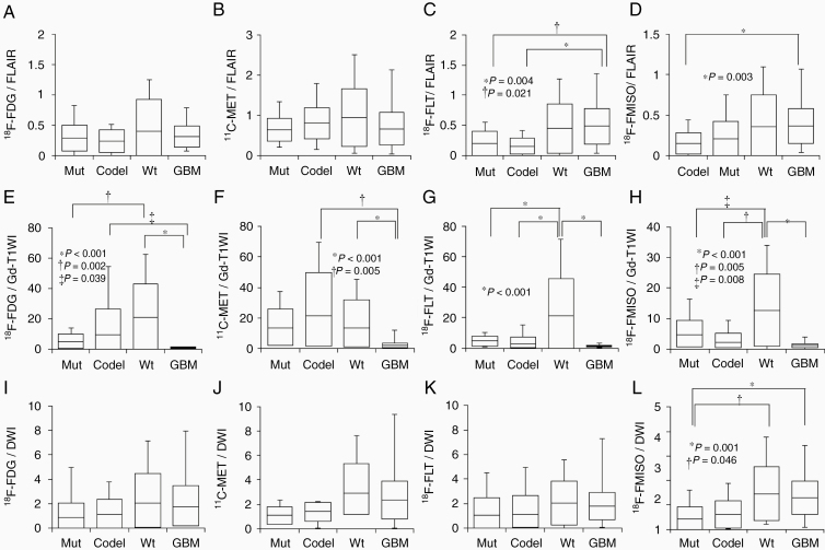
Figure 3.
Box plots indicating the comparisons between the FLAIR volume and the MTVs of 18F-FDG (A), 11C-MET (B), 18F-FLT (C), and 18F-FMISO (D), between the Gd-T1WI volume and the MTVs of 18F-FDG (E), 11C-MET (F), 18F-FLT (G), and 18F-FMISO (H), and between the DWI volume and the MTVs of 18F-FDG (I), 11C-MET (J), 18F-FLT (K), and 18F-FMISO (L) for each of the 4 glioma subtypes. Lines within the boxes indicate the average, boxes represent standard deviation, and whiskers denote minimum–maximum.

