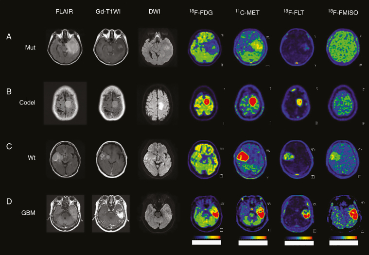
Figure 4.
MRI (FLAIR, Gd-T1WI, and DWI) and PET images (18F-FDG, 11C-MET, 18F-FLT, and 18F-FMISO) in representative glioma patients. (A) A 29-year-old female patient in Mut subtype with the accumulation of 11C-MET and 18F-FLT. (B) A 38-year-old male patient in Codel subtype with high accumulation of 11C-MET, and slight accumulation of 18F-FLT. (C) A 58-year-old male patient in Wt subtype with the accumulation of 11C-MET and higher accumulation of 18F-FLT and 18F-FMISO than Mut. (D) A 69-year-old male patient with GBM with the highest accumulation of 11C-MET, 18F-FLT, and 18F-FMISO.

