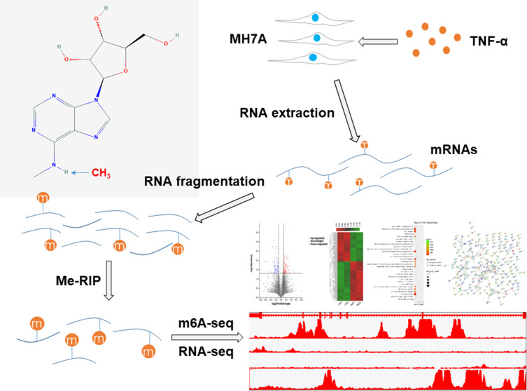
Figure 1.
A schematic diagram of m6A-seq and RNA-seq analyses of MH7A cells. MH7A cells were stimulated with TNF-α, and total RNA was extracted from MH7A cells. Then, the RNA was fragmented, and m6A RNA was isolated as described by the m6A-RNA immunoprecipitation (Me-RIP) assay. The m6A-seq library and RNA-seq library of these mRNAs were constructed, and then sequenced for analysis.

