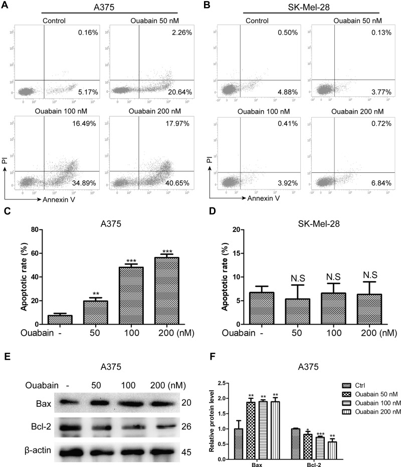
Figure 3.
The effects of ouabain on apoptosis induction in A375 and SK-Mel-28 cells. Melanoma cells were treated with indicated concentrations of ouabain for 48 hours. The samples of A375 (A) and SK-Mel-28 (B) cells were stained by Annexin V/PI, the induction of apoptosis was detected by flow cytometry. The histograms of apoptotic rate in A375 (C) and SK-Mel-28 (D) cells are shown, which consisted of early apoptotic rate and late apoptotic rate. (E). The expression of bcl-2 and bax were determined by Western blotting analysis in A375 cells. (F). The histograms showed the relative protein level of each group compared with control. The results are shown as mean±SD by Student’s t-test (two-tailed). NS, no significant difference, *P<0.05, **P<0.01, and ***P<0.001.

