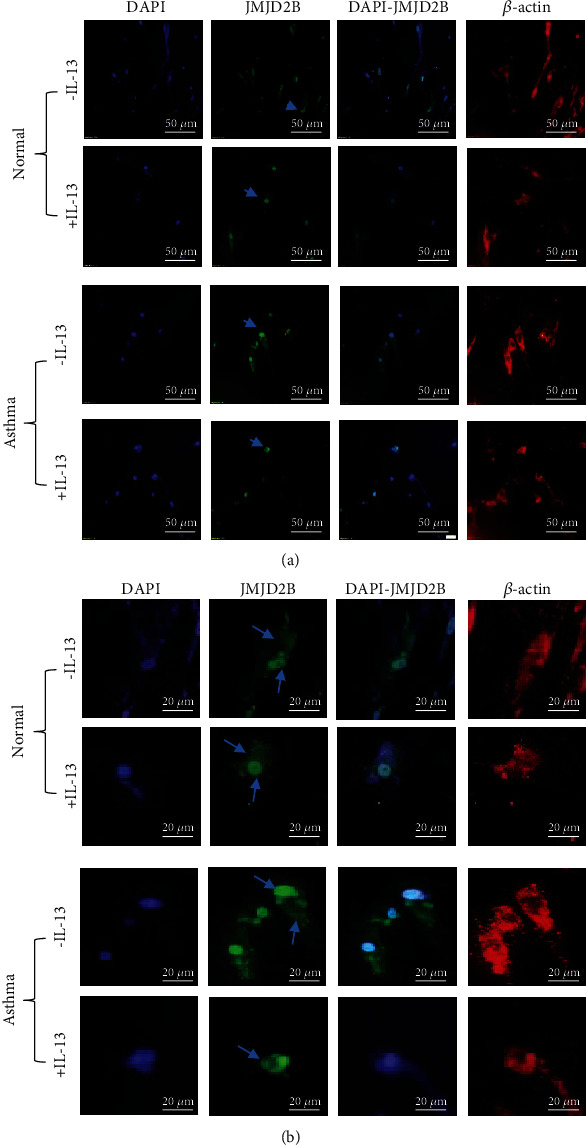
Figure 4.

JMJD2B subcellular localization after IL-13 stimulation in normal and asthmatic fibroblasts. Immunofluorescence staining of in normal and asthmatic fibroblasts stimulated with IL-13 that were stained for DNA (DAPI; blue), JMJD2B (green), β-actin (red). The images were observed under a microscope at 20x magnification (a) and at 40x magnification (b). Arrows indicate subcellular localization of JMJD2B in fibroblasts.
