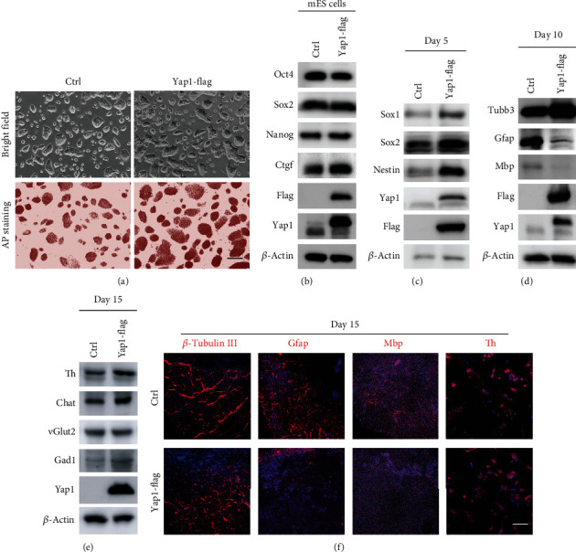
Figure 4.

Yap1 overexpression in mESCs phenocopied the glial cell lineage defect differentiated from Lin28a/b OE cells. (a) Phase-contrast microscopy and AP staining of Ctrl and Yap1 constitutively expressed (Yap1 OE) mouse ESCs grown under 2i + LIF medium. Scale bar, 200 μm. (b) Western blot analyses of total proteins from Ctrl and Yap1 OE mouse ESCs using the indicated antibodies. Oct4, Sox2, and Nanog are pluripotent stem cell markers. (c) Western blot analyses of total proteins from Ctrl and Yap1 OE mouse ESC differentiated cells on day 5 using the indicated neural stem cell markers: Sox1, Nestin, and Sox2. (d) Western blot analyses of total proteins from Ctrl and Yap1 OE mouse ESC differentiated cells on day 10 using the indicated neuronal marker: β-tubulin III, glial markers: Gfap and Mbp. (e) Western blot analyses of total proteins from Ctrl and Yap1 OE mouse ESC differentiated cells on day 15 using the indicated neuronal subtype markers: Th, vGlut2, Gad1, and Chat. (f) Immunofluorescence staining of the Ctrl and Yap1 OE mouse ESC differentiated cells on day 15 using the neuronal marker β-tubulin III, glial markers Gfap and Mbp, and neuronal subtype markers Th. Cell nuclear was stained with DAPI. Scale bar, 200 μm.
