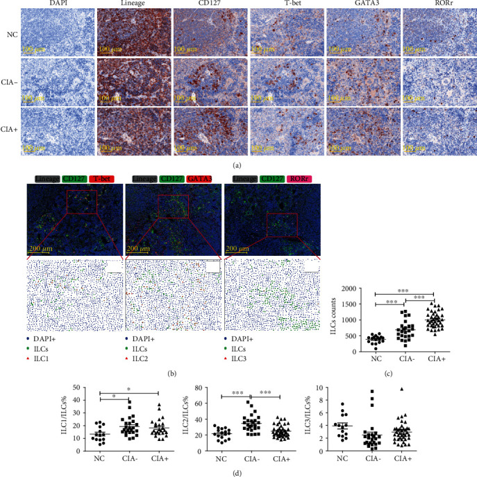
Figure 4.

ILC subsets in the spleens of mice with CIA and NCs. (a) Extracted single spectrum of each labeled marker from immunofluorescence staining shown as an immunohistochemistry image. (b) Multiple fluorescence image (top) and phenotyping analysis by HALO software (bottom), in which each symbol represents a phenotype (blue dots: total cells (DAPI+); green dots: total ILCs (Lineage-CD127+); red triangle (left): ILC1s (Lineage-CD127+T-bet+), red triangle (middle): ILC2s (Lineage-CD127+GATA3+); red triangle (right): ILC3s (Lineage-CD127+RORγt+)). (c) Numbers of total ILCs in each field of view. (d) Proportions of ILC1s, ILC2s, and ILC3s among total ILCs. Each point represents a random observation from a spleen section from 2 to approximately 4 mice per group. NC: n = 17; CIA−: n = 25; and CIA+: n = 38. NC: negative control; CIA: collagen-induced arthritis. Data are expressed as means ± SDs and analyzed using the unpaired Student's t-test. Spearman's rank correlation coefficients and p values are indicated. ∗p < 0.05, ∗∗∗p < 0.001.
