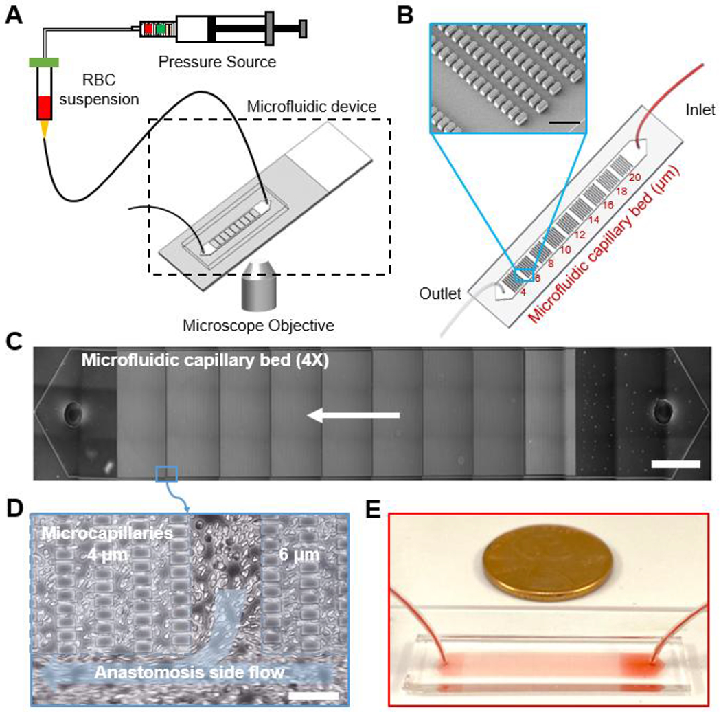
Figure 1. The OcclusionChip microfluidic platform allows standardized assessment of RBC mediated microcapillary occlusion.

(A) A schematic view of the assay is shown. RBCs diluted in PBS at 20% hematocrit are infused into the microchannel using a manual syringe pump at constant pressure. (B) A schematic of the OcclusionChip design is shown. The OcclusionChip features nine micropillar arrays with microcapillaries ranging from 20 to 4 μm, coupled with two anastomosis-mimicking side pathways that are 60 μm wide. Inset: Scanning electron microscopy image showing the microvascular features. Scale bar represents a length of 50 μm. (C) The entire microchannel is shown at 4X magnification. Arrow indicates flow direction. Scale bar represents a length of 2 mm. (D) Close-up view showing the anastomosis side flow. Scale bar represents a length of 50 μm. (E) Macro view of the OcclusionChip with a blood sample is shown.
