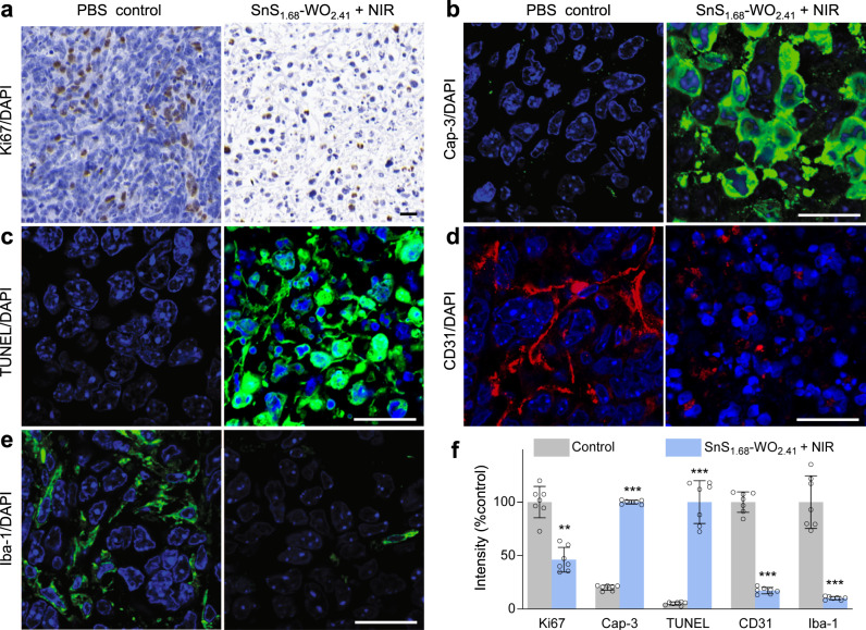
Fig. 6. The change of TME after hole/hydrogen therapy with the SnS1.68–WO2.41 nanocatalyst.
Immunohistochemical analysis of Ki67+ proliferating cells (a), caspase-3+ apoptotic cells (b), TUNEL+ apoptotic cells (c), CD31+ tumor vessels (d), and Iba1+ tumor-associated macrophages (e) in 4T1 tumors, and corresponding quantification results (n = 7 biologically independent samples) (f). All the scale bars, 20 μm. P values were calculated by the two-tailed Student’s t-test (**P = 0.000006, ***P < 0.000001). Data are presented as mean values ± SD.

