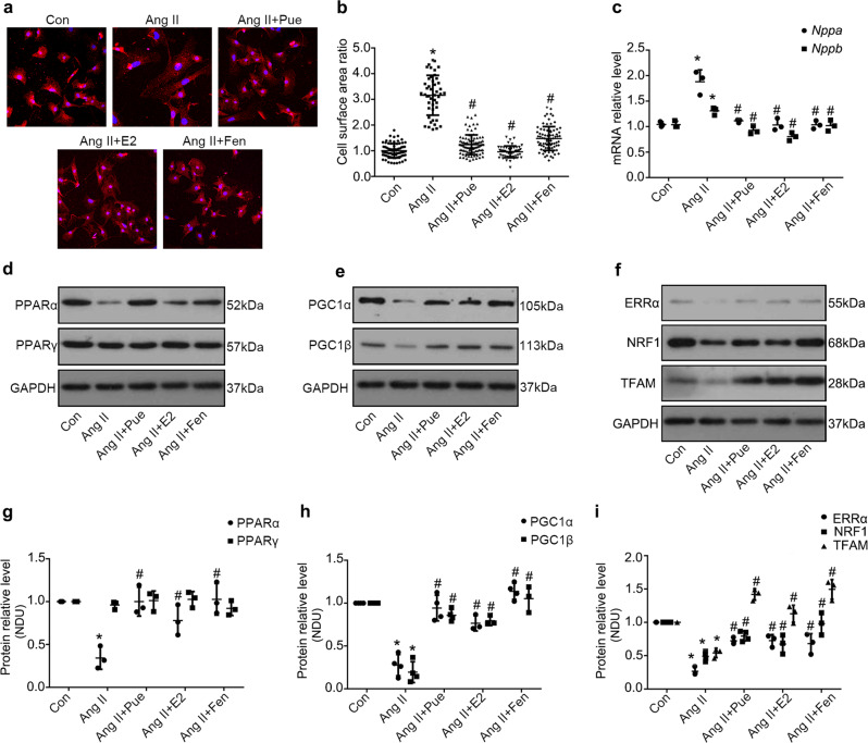
Fig. 5.
Puerarin upregulates the PPARα/PGC-1 pathway in Ang II-induced cardiomyocyte hypertrophy. a, b: Representative confocal images (a) and measurement of surface area (b) of cardiomyocytes subjected to different treatments, as indicated; n = 40–50 cells per group. c Reverse transcription-polymerase chain reaction analysis of atrial natriuretic peptide A (Nppa) and natriuretic peptide B (Nppb) in NRCMs subjected to the given treatments. Data are representative of three independent experiments. d, g Representative Western blots of whole lysates (d) and fold increases in the relative densitometric values of PPARα and PPARγ (g) in each group. e and h: Representative Western blots of whole lysates (e) and fold increases in the relative densitometric values of PGC-1α and PGC-1β (h) in each group. f, i Representative Western blots of whole lysates (f) and fold increases in the relative densitometric values of estrogen-related receptor alpha (ERRα), nuclear respiratory factor 1 (NRF1), and mitochondrial transcription factor A (TFAM) (i) in each group. Data are presented as the mean ± SD. Statistical significance was determined using one-way ANOVA coupled with Tukey’s multiple comparison post hoc test. *P < 0.05 vs. Con group; #P < 0.05 vs. Ang II group; representative Western blots and the densitometric analysis from three or four independent experiments

