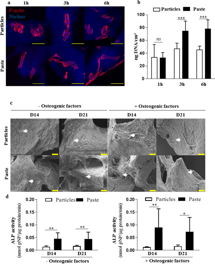
Figure 1.
Attachment and osteoblastic commitment of hBM-MSCs cultured in contact with the particular bone graft or the bone paste. (a) Representative confocal images of stained hBM-MSCs. Red (phalloidin-AF568): F-actin, blue (Hoechst): nucleus. The particular bone graft and the bone paste were visible as a result of their blue auto fluorescence. Scale bars: 100 µm. (b) Quantification of the total DNA in hBM-MSCs in contact with the particular bone graft or the bone paste. (c,d) SEM images and ALP activity in lysates of hBM-MSCs grown in contact with the particular bone graft or bone paste with or without osteogenic factors for 14 and 21 days. Scale bars: 20 µm. The results are expressed as means ± the SD (N = 3, n = 3; *p < 0.05, **p < 0.01 ***p < 0.001).

