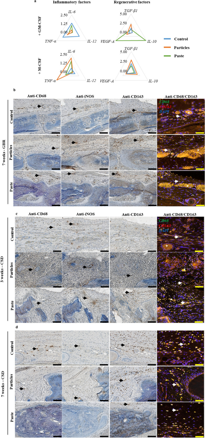
Figure 6.

Effect of the particular bone graft and the bone paste on monocyte/macrophage polarization. (a) mRNA expression in human monocytes cultured in the presence of GM-CSF or M-CSF for 3 days in contact with the particular bone graft or the bone paste. The Y-axis represents the mean relative mRNA expression level of the specified genes (N = 3). Representative immunohistochemical and immunofluorescent stainings on successive undecalcified 7-µm thick sections, (b) in the GBR model after 7 weeks, and (c) in the CSD model after 3 and 7 weeks. Immunohistochemical stainings were performed for CD68, iNOS, CD163, and immunofluorescence with double labeling of CD68 (green)/CD163 (red) and DAPI (blue) for the nuclei. For single labeling, positive cells were stained with a brown precipitate (black arrow), for double labeling, double-positive cells appeared as light yellow (white arrow). Typical auto fluorescent red blood cells were visible (white stars). Scale bars: 100 µm for the immunohistochemistry, 50 µm for the immunofluorescence images.
