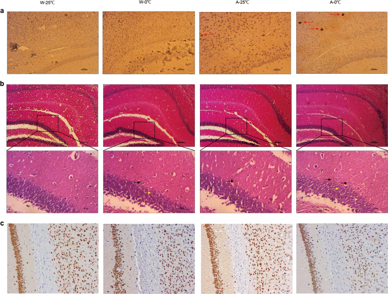
Fig. 6. Effect of the daily administration of 0 °C IW on amyloid lesions and neuropathy in the cortices and hippocampi of APP/PS1 mice.
After the wild-type C57BL/6 mice (W) and APP/PS1 mice (AD model, A) were administered intragastric water (IW, 10 mL/kg, 2 times/day) at either 0 °C (the W-0 °C and A-0 °C groups) or 25 °C (the W-25 °C and A-25 °C groups) for 35 consecutive days, amyloid lesions and neuropathy in the cortex and hippocampus were observed by immunohistochemistry and H&E staining, respectively. a Yellowish-brown or black-brown senile plaques (red arrow) in the cortex and hippocampus of each mouse were counted (×200). b Lesions in the cortex and hippocampus were observed, and lesions with scattered nerve cells (green arrow) and enlarged gaps (blue arrow) in the CA3 region of the hippocampus are shown (×100; magnification: ×400). c Representative NeuN staining of cells in each group. Brown or black-brown cells are NeuN-positive cells (×200).

