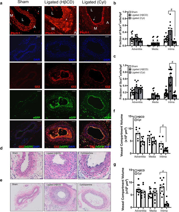
Fig. 4. Inhibition of Hh signalling attenuates intimal thickening and Sca1 cell expansion following iatrogenic flow restriction.
a Representative immunofluorescence images of Hh receptor patched 1 (Ptch1) (red), Hh target gene Gli2 (red), Sca1-eGFP (green) expression and merged (eGFP/Gli2/DAPI) in carotid artery cross sections from sham-operated, ligated vehicle control (HβCD), and ligated + cyclopamine (10 mg/kg, IP) treated mice after 14 days. Scale bar = top panel: 50 µm, all other panels: 20 µm. b, c Cumulative data showing the fraction of Sca1-eGFP cells within the adventitia, media and intima from sham-operated, ligated vehicle control (HβCD), and ligated + cyclopamine (Cyl) Sca1-eGFP mice after b 7 days and c 14 days. Data are the mean ± SEM of five sections per experimental group, #p ≤ 0.05 vs ligated HβCD controls and are representative of a minimum of four animals per experimental group. d, e Representative H&E staining of carotid artery cross sections from sham, ligated control (HβCD vehicle), and ligated + cyclopamine (Cyl) treated animals after 7 d (d) and e 14 d. Scale bar = 50 µm. f, g Morphometric analysis of vessel compartment volumes for ligated control (HβCD), and ligated + cyclopamine (Cyl) treated animals after f 7 d and g 14 d. Data mean ± SEM of 7–12 sections analysed from four animals, #p ≤ 0.05 vs ligated control (HβCD).

