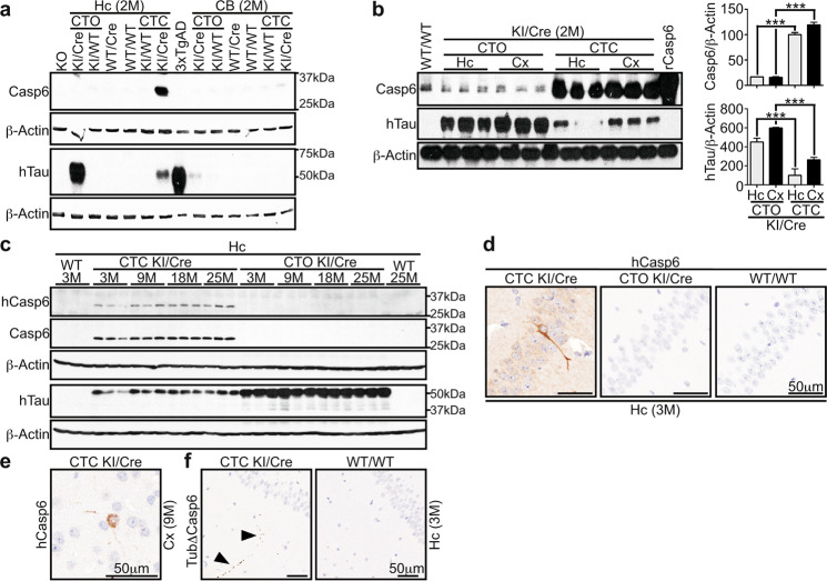
Fig. 1. Specific co-expression of active hCasp6 with hTau in the cortex and hippocampus of CTC KI/Cre mice.
Western blot with 15 μg total proteins from (a, b) 2 month (M) old and (c) 3–25-month-old CTO KI/Cre, CTO KI/WT, WT/Cre, WT/WT, CTC KI/WT, or CTC KI/Cre hippocampus (Hc), cerebellum (CB), or cortex (Cx) with anti-Casp6, anti-hTau, anti-hCasp6, and anti-β-Actin antibodies. In a, proteins from Casp6 KO hippocampus or Tau KO brainstem (KO) were used as negative controls for Casp6 and Tau analyses, respectively. Cx proteins from 3xTgAD were used as positive control for hTau. In b, the histograms represent densitometric analyses of the western blot shown. Data are expressed as ratios of Casp6 or hTau over β-Actin, relative to CTC KI/Cre hippocampus (n = 3/genotype/structure). One-way ANOVA followed by Bonferroni’s post-hoc test was performed. ***p < 0.001. d-f Representative micrographs from n = 5/genotype/structure/age/antibody for 3 M hippocampus (d) and 9 M cortex (e) with anti-hCasp6, and 3 M hippocampus with anti-Tub∆Casp6 (f).

