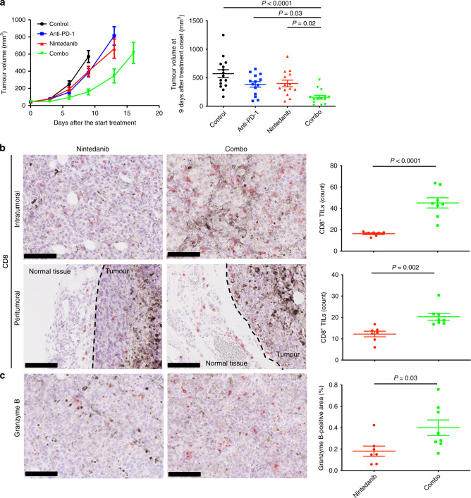
Fig. 5. The combination of nintedanib and PD-1 blockade shows an increased antitumour efficacy.
a Time course of tumour volume (left) as well as tumour volume at 9 days after treatment initiation (right) for subcutaneous B16-F10 tumours treated with anti–PD-1, nintedanib or the combination of both agents. Data are means ± SEM for 14 or 15 mice in each arm (pooled from two independent experiments). P values were determined by one-way ANOVA with Tukey’s correction for multiple comparisons and are shown only if <0.05. b Representative immunohistochemical staining of CD8 (left) as well as the number of CD8+ TILs (right) for intra- and peritumoural regions of B16-F10 tumours derived from mice treated with nintedanib or the combination of anti–PD-1 and nintedanib for 7 days. Scale bars, 100 μm. c Representative immunohistochemical staining (left) as well as the percentage positive area (right) for granzyme B in B16-F10 tumours derived from mice treated with nintedanib or the combination of anti–PD-1 and nintedanib for 7 days. Scale bars, 100 μm. Quantitative data in b and c are means ± SEM for seven or eight mice per group. P values in b and c were determined with the unpaired t-test.

