Figure 2.
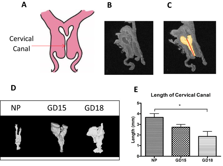
Generating 3D models of the murine cervical canal for accurate measurements of its length. (A) A schematic of murine cervical anatomy presented in the frontal plane, with the opening in the middle indicating the cervical canal. (B) A representative anatomical scan of the non-pregnant murine cervix acquired by T2-weighted imaging using a 7-T scanner (C) 3D models of the cervical canal (shown in yellow) were generated by the software Amira (Thermo Fisher Scientific, version 6.4.0) and were used to measure length of cervical canal. (D) Representative 3D models of non-pregnant (NP), and pregnant cervices on gestational day (GD)15 and GD18 show a transition from a Y shape to a U shape. (E) Data is presented as mean ± SEM, showing that the length of the cervical canal (measured from ectocervix to immediately before the bifurcation of the endocervix) significantly decreases at late gestation (GD18, 2.09 mm ± 0.45) as compared to NP mice (3.66 mm ± 0.35). Statistical significance was determined by one-way ANOVA with Bonferroni post-hoc test. *p < 0.05 (n = 3–6/GD).

