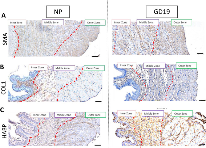
Figure 9.
Smooth muscle actin (SMA), collagen type 1 (COL1) and hyaluronic acid binding protein (HABP) immunostaining within transverse sections of the non-pregnant and pregnant murine endocervix. Brown deposit indicate positive immunostaining. Blue shows hematoxylin counterstaining. Three zones are identified in both NP and GD19 samples: inner, middle and outer. (A) Staining of SMA is absent in the inner zone, while prominent in a middle zone (circular muscle fibers), and in an outer zone (longitudinal muscle fibers); (B) Staining of COL1 is localized in the sub-epithelial region of the inner zone specifically in the NP sample, but not in GD19 sample. (C) HABP shows similar weak staining throughout all three zones in NP endocervix, but the immunostaining is more intense in GD19 sample showing no heterogeneity. Magnification is at 200×. Scale bar = 100 μm.

