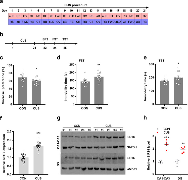
Fig. 1.
Chronic stress leads to an increase in mRNA and protein level of hippocampal SIRT6. a Time course and design of CUS. b Experimental timeline is shown for in vivo CUS-induced mice model. c–e SPT, FST, and TST were performed on the 21th day. The data are expressed as mean ± SEM (n = 12 per group). *P < 0.05, **P < 0.01 compared with CON. f Quantitative real-time PCR analysis of SIRT6 mRNA expression in the hippocampus. The data are expressed as mean ± SEM (n = 12 per group). ***P < 0.001 compared with CON. g Western blotting analysis showed that CUS increased the protein level of SIRT6 in hippocampal CA1-CA3 and DG area. h Quantitative analysis of SIRT6 protein level using Image J software. The data are expressed as mean ± SEM (n = 3 per group). ***P < 0.001 compared with CON

