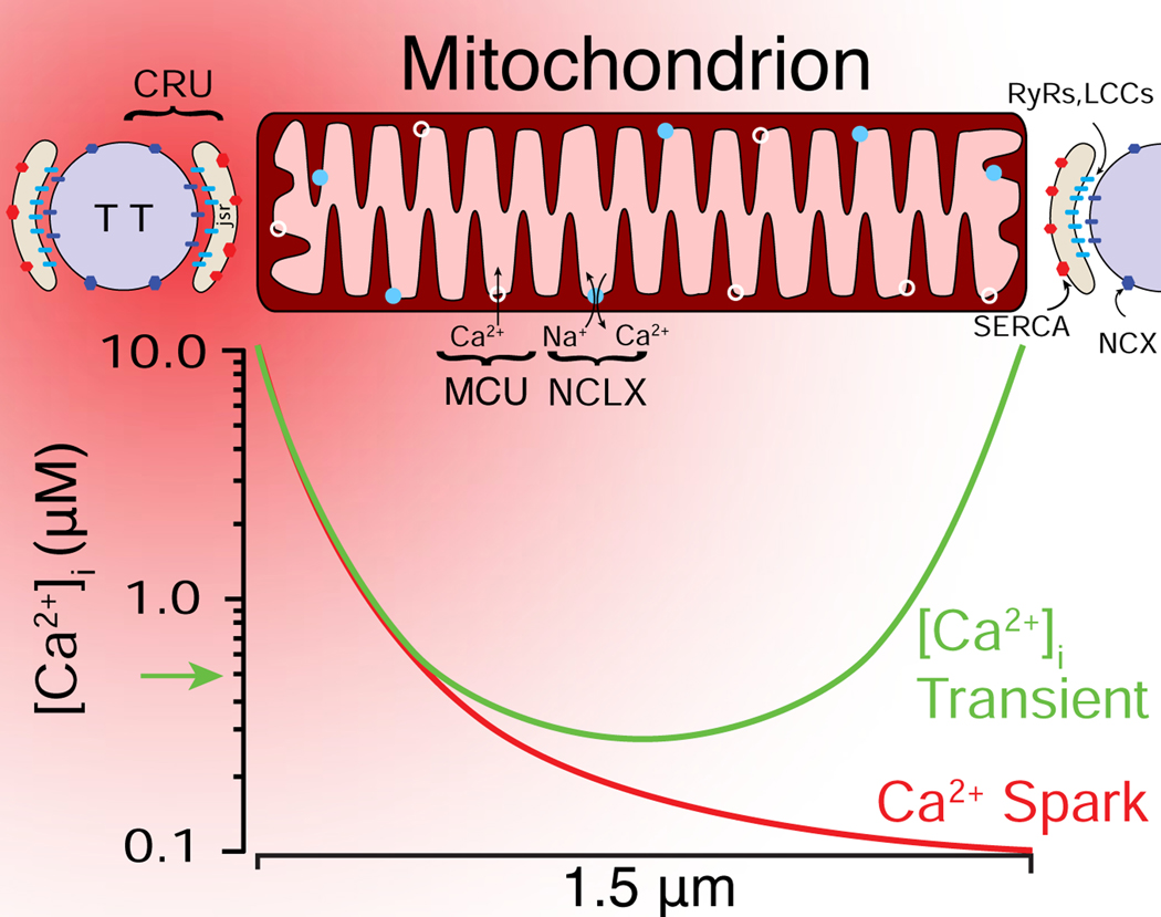
Figure 2. spatial distribution of cardiac mitochondrial Ca2+ signaling components.
A spatial representation of a Ca2+ spark (red gradient) initiated at the Ca2+ release unit (CRU), which is located between the transverse-tubule (TT) and the junctional SR (JSR) membranes. At the peak of a Ca2+ spark, [Ca2+]i briefly (10 ms) bathes the end of a mitochondrion with high [Ca2+]i (5–10 μM). During a [Ca2+]i transient, multiple CRUs release Ca2+, bathing both ends of the mitochondrion with high [Ca2+]i. LCCs, L-type Ca2+ channels; RyR2s (ryanodine receptor type 2); SERCA: sarcoplasmic reticulum and endoplasmic reticulum Ca2+ ATPase; NCX: Na+-Ca2+ exchanger; MCU: mitochondrial Ca2+ uniporter; NCLX: mitochondrial NCX. Adapted from [46].

