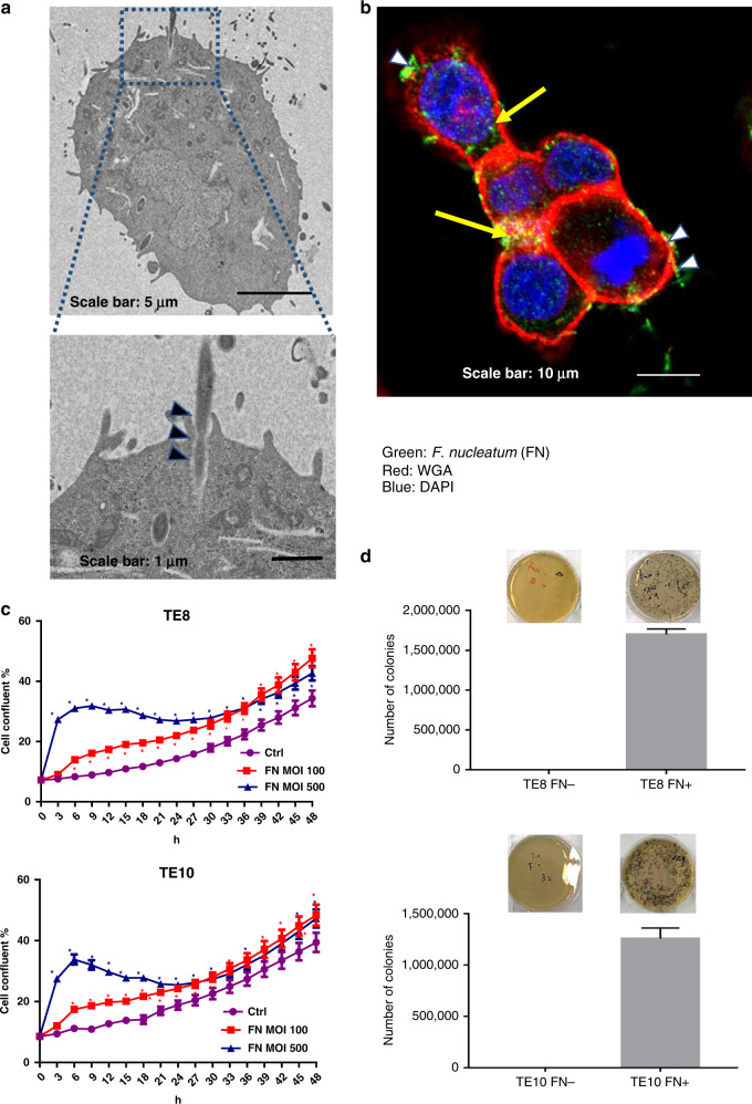
Fig. 2. F. nucleatum invades and survives in ESCC cells.
a F. nucleatum invades into the cancer cell as observed by transmission electron microscopy (17,500 ×3 magnification) in TE10 cell. Scale bar, 1 µm. b LSCM analysis of F. nucleatum adhered and invading into the TE10 cells. Cytomembrane (red), cell nucleus (blue), and F. nucleatum (green). Black arrowheads indicate invasion in a, and white arrowheads indicate adherence in b. c Proliferation assay by Incucyte, with or without F. nucleatum, for 48 h at MOI 100 and 500 in TE8 cells and TE10 cells. *P < 0.05 (red and blue) indicates significant difference between the MOI 100 and ctrl groups and between the MOI 500 and ctrl groups. d F. nucleatum invasion in TE8 and TE10 cancer cell lines. Growth of intracellular F. nucleatum into colonies on the GAM agar.

