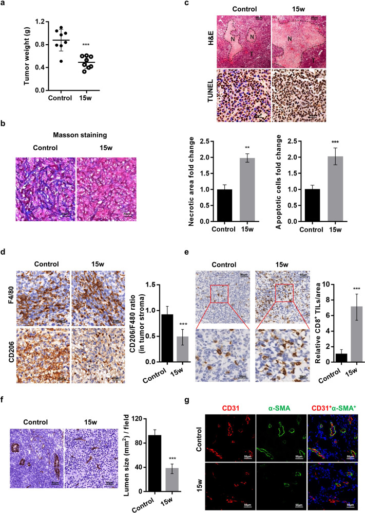
Fig. 6. SMS2 inhibitor attenuates tumor growth in a 4T1 breast cancer model.
a Weights of the 4T1 primary breast tumors from treated mice. b Collagen staining in tumor tissues. c H&E staining of tumor sections showing intensely stained necrotic areas (N necrotic tissue, T tumor tissue) and TUNEL staining for apoptotic cells. d Tumor IHC staining and quantification of the CD206 to F4/80 ratio to indicate the level of M2 macrophages. e Representative IHC staining of tumor-infiltrating CD8+ lymphocytes (×100 magnification and enlarged ×400 magnification). Quantifications of normalized CD8+ lymphocytes from five fields per tumor are shown. The lumen size of the tumor vessels was analyzed by CD31 IHC (f) and CD31 and α-SMA dual IF staining (g). The results are shown as the mean ± SD. **P < 0.01; ***P < 0.001 (n = 8 mice/group).

