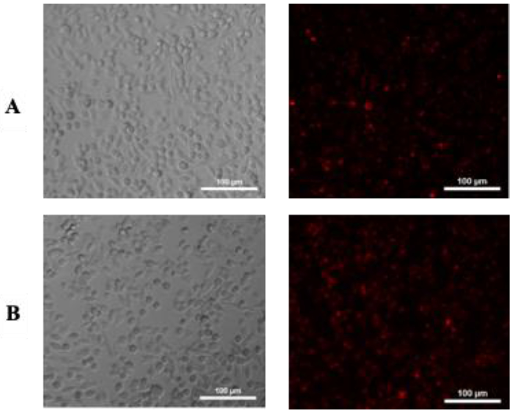
Figure 6.

Internalization of lipid formulations by B16F10 cells determined by fluorescence microscopy. From the left to right: Bright field and lipid formulations internalized. A) images of B16F10 cells treated with (1-DSPE-PEG) and B) (1-DSPE-PEG/KI), respectively, after 4 h of incubation.
